A healthy dog’s gums should be a vibrant bubblegum pink or a soft rosy hue; when pressed, they should briefly turn white for 1–2 seconds before quickly returning to their original color. If the gums turn pale, deep red, purple, or yellow—or if mottled black patches suddenly appear—it may indicate that your dog is suffering from an underlying medical condition, ranging from severe dehydration to life-threatening sepsis.
In this comprehensive guide, we’re breaking down the exact visual cues that separate healthy gums from unhealthy gums during a medical emergency. Plus, you’ll learn how to run a quick, 2-second CRT (Capillary Refill Time) test right at your kitchen table—a simple home check that can help you screen for sudden shock and sepsis before it’s too late.”
📋 Table of Contents
| Gum Color | Risk Level | Potential Condition | Pet Parent Action Guide |
|---|---|---|---|
| 🌸 Bubblegum Pink | 🟢 Safe | Healthy blood flow, optimal oxygen levels, and robust circulation. | Maintain daily teeth brushing and routine oral care. |
| 🏳️ Pale / Paper White | 🔴 Critical | Severe anemia, shock, or life-threatening internal bleeding. | Seek emergency care immediately! No safe home remedies. |
| 🟨 Yellow / Jaundice | 🔴 Critical | Liver disease, bile duct obstruction, or acute hemolysis (toxin poisoning). | Seek immediate veterinary care! Requires urgent blood tests and ultrasounds. |
| 🟦 Blue / Purple (Cyanotic) | 🚨 Life-Threatening | Severe oxygen deprivation (advanced heart disease, choking, or suffocation). | Absolute emergency! Rush to the vet immediately for professional oxygen therapy[cite: 4]. |
| 🟥 Bright / Brick Red | 🔴 Critical | Severe systemic infection (sepsis), heat stroke, or severe local gum inflammation. | Go to the vet immediately! For heat stroke, use wet towels to cool them down during transit[cite: 4]. |
| 🍂 Dry / Sticky Pink | 🟡 Warning | Severe dehydration causing decreased blood volume and sticky mucous membranes. | Do not force water down their throat. Seek veterinary help for safe IV fluid replacement. |
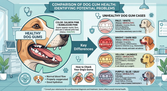
To help pet owners establish a scientifically sound basis for assessment, we have consulted the AAHA (American Animal Hospital Association) Canine Life Stage Health Assessment Guidelines. We have compiled the various states of canine gum color, tactile indicators, and potential clinical warning signs into the quick-reference chart below. I recommend that every pet owner take a screenshot of this chart and save it to serve as the first line of defense for your pet’s health right at home:
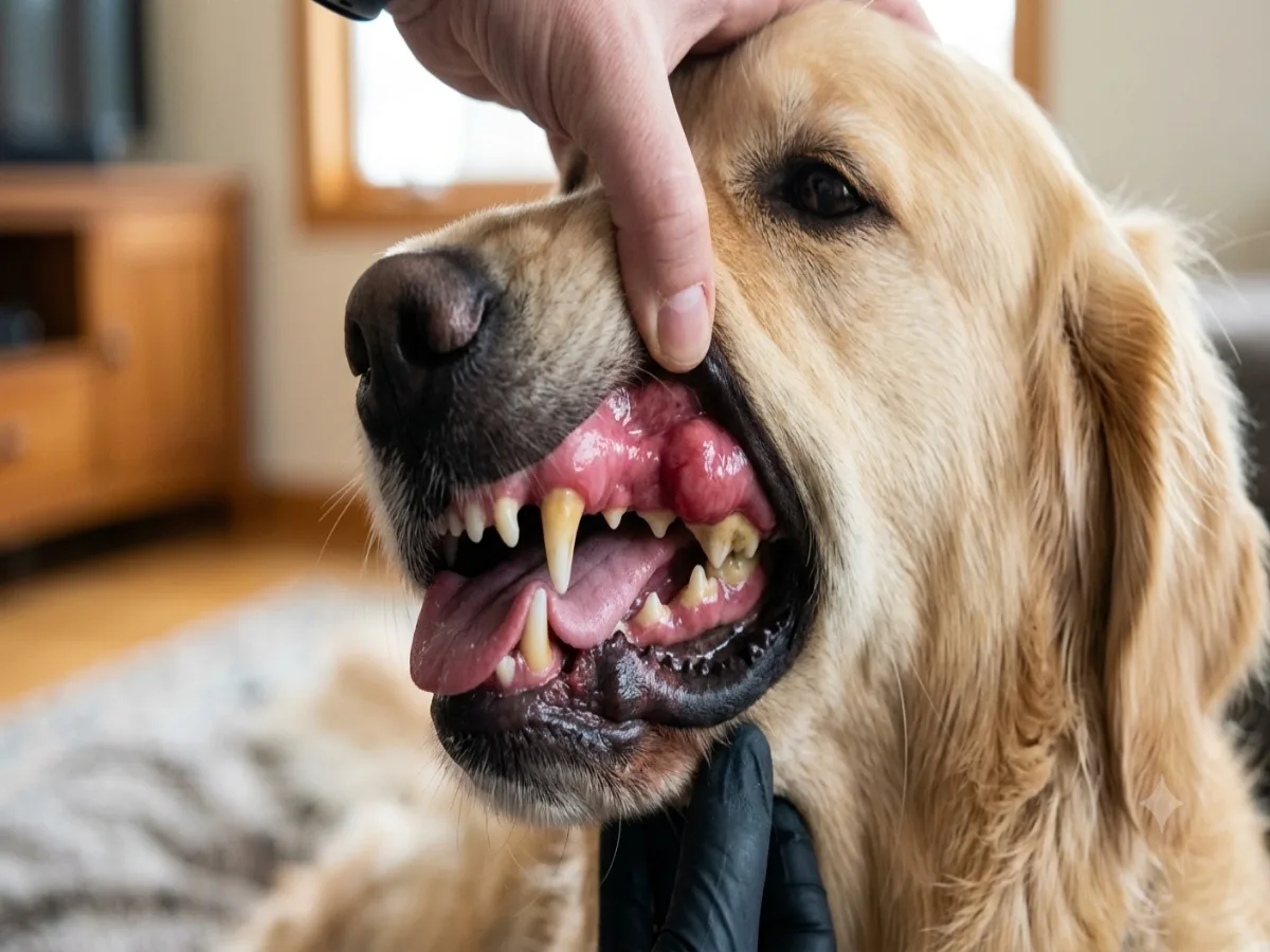
The Gold Standard: Salmon-Pink Dog Gums (Healthy)
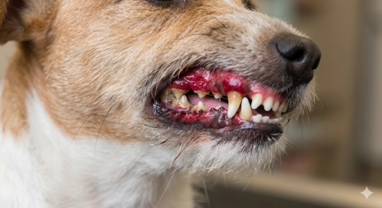
If you are dealing with sick dog gums that appear washed-out—having lost their natural flush and presenting as a very pale pink, or even a lifeless, stark white—this is a red flag of a serious underlying problem.

Black gums in dogs can be either a normal occurrence or a warning sign of a potentially dangerous condition. When it comes to spotting black patches in your dog’s mouth, you need to play detective. Ask yourself two crucial questions: Did this spot show up overnight, or has it always been there? and Is the spot flat, or is it raised and bumpy?
If your dog has had black spots on its gums since puppyhood—and as it ages, the entire gum tissue darkens uniformly while remaining flat, smooth, and moist—this is typically considered normal.
If your dog’s gums are typically a healthy pink, but have recently and suddenly developed localized black patches, this requires your immediate and close attention.
According to statistics from the ACVIM (American College of Veterinary Internal Medicine) and numerous studies on canine oral oncology, malignant melanoma is the most common oral malignancy in dogs, accounting for 30% to 40% of all canine oral cancer cases.
This tumor is highly aggressive; not only does it rapidly destroy the alveolar bone within the oral cavity, but it also metastasizes very easily—even at an early stage—to the lungs or regional lymph nodes via the lymphatic system.
Melanoma is a master of disguise. When it first appears, it may look like nothing more than a tiny black speck lodged between the teeth—resembling a bit of food stuck in the gums after eating meat, or a minor injury sustained from chewing on a stick.
As pet owners, we must never rely solely on the naked eye to determine whether a newly appeared black growth is benign or malignant. If, while brushing your dog’s teeth or performing a routine check, you discover any new, raised, firm-to-the-touch, or easily bleeding black tissue, the only scientifically sound course of action is to take your dog to a veterinary clinic immediately. A veterinarian will typically perform a Fine Needle Aspiration (FNA) or a tissue biopsy to rule out this potentially fatal “black gum crisis” for both you and your furry companion.
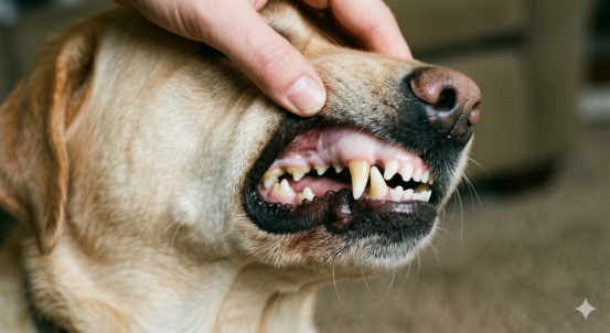
In veterinary medicine, the gums are often the very first area a veterinarian examines during an emergency. This is because when a dog suffers severe trauma, infection, or faces a life-threatening situation, the body activates a “blood redistribution” mechanism as a self-preservation measure—diverting blood flow away from peripheral organs (such as the gums and skin) to prioritize and safeguard the heart and brain.
Therefore, whenever you suspect your dog’s life is in danger, simply lifting their lips to check their gums serves as the quickest method for crisis screening.
When pet owners anxiously type “early signs of canine sepsis” into search engines, they are often looking for a confirmation method that allows for immediate action. Clinically, the gold standard self-test recommended by veterinarians for home use is the CRT test (Capillary Refill Time test).
This test assesses a dog’s cardiovascular function, perfusion status, and the potential presence of a severe systemic infection (sepsis/septicemia).
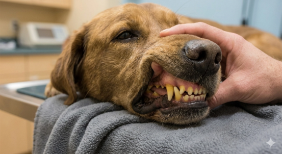
Gently lift your dog’s upper lip, taking care to avoid any areas of natural pigmentation (dark spots). Using the pad of your thumb, apply firm pressure to a patch of healthy, pink gum tissue and hold for 2 seconds. During this time, the blood within the local capillaries will be temporarily displaced by the pressure.
Abruptly release your thumb. You will observe that the specific spot you just pressed has turned a distinct shade of white due to the temporary lack of blood flow. At this moment, watch your stopwatch or count silently to yourself (e.g., “one second… two seconds…”).
Observe how many seconds it takes for the whitened patch of gum tissue to return completely to its original pink color, matching the surrounding tissue.
Normal Status (1–2 seconds):
he gums rapidly return to a pink color within 1 to 2 seconds. This indicates that the dog’s heart is pumping blood vigorously, blood pressure is normal, and systemic microcirculation perfusion is good.
Abnormally Slow (Over 2 seconds):
If it takes longer than 2 seconds—or even 3 to 4 seconds—for the gums to sluggishly regain their color, it suggests that the dog may be suffering from hypovolemic shock, internal bleeding, severe dehydration, or heart failure.
Abnormally Fast (Instantaneous Return / Brick-Red):
The moment you release your finger—in less than 0.5 seconds—the gums become intensely engorged with blood, and the entire oral mucosa takes on a striking brick-red hue. This is the most classic sign of the early compensatory phase of systemic sepsis (blood poisoning)! At this stage, pathogenic bacteria release large quantities of endotoxins, causing severe dilation and congestion of systemic microvessels. If observed, immediate veterinary attention is mandatory.
The information presented herein—including descriptions of canine gum coloration, chart data, at-home CRT (Capillary Refill Time) testing procedures, and critical warning signs—is derived from authoritative veterinary medical guidelines and the author’s personal experience as a pet owner. Its sole purpose is to assist pet owners in maintaining a proactive awareness of their pets’ daily health status. Given that every dog possesses a unique constitution, age, and medical history, changes in oral coloration may be the result of the complex interplay of various systemic diseases. Consequently, the content of this article does not constitute a formal clinical diagnosis, treatment recommendation, or emergency medical prescription.